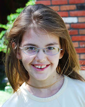
...pretty good after spending another 6 hours in impromptu appointments at Primary Children's (and none of that included time to eat lunch!). We got to mosey around 4 separate clinics on different floors and were even told that we know how to navigate the hospital better than some of the employees - how'd that happen?
We got to the Imaging waiting room right at 10:30 and they called Sada in 2 minutes later to start her MRI. She didn't get music or a movie this time, but she did get the super expensive REALLY powerful scanner. Comparing the two machines they have, these latest pictures look like professional portraits next to cell phone photos (the previous scans). We didn't get to bring home the dvd of the photos because their disk writing machine froze this morning, but they are going to mail it to us this week (so I'll put them on the blog soon).
The scan showed:
1. The little spot on the brain stem where the tumor had infiltrated hasn't changed at all and wasn't showing up as anything on the new MRI. That means it probably won't come back.
2. The little "artifact" (thingy) that appeared after surgery at the base of the cerebellum hasn't changed - so it is most likely part of an artery or a calcification - but it's not any cause for concern because it hasn't changed a bit.
3. The cerebellar area at the top of the tumor (where the surgeon said it was really hard to get into without damaging healthy tissue) does show up lighter with the contrast MRIs, so the tumor cells have been growing there. It's about the size of a pinkie nail right now. Comparing it with the post-op MRI, the tumor spot is a lot clearer and larger... mainly because there was swelling and some bleeding obstructing the view the day after surgery. It is sitting below the 4th ventricle on the cerebellum and has enough room to grow quite a bit before it would start causing symptoms like gross central balance loss.
We talked to the neurosurgeon, he wants to wait another 2-3 months for another MRI and see what happens to area #3 - 1 out of 3 resected (surgically removed) JPAs grows more, 1 out of 3 stays the same forever, and 1 out of 3 goes away on it's own. But he did send us up for an oncology consult so he could have someone else collaborate his opinion. The doctor completely agreed with a wait-and-see approach, and if it does grow a lot over the next few months she'd much rather do surgery than chemo or radiation. Yeah, we'll agree with that opinion. Believe it or not, surgery is much less traumatic than chemo or radiation... and doesn't carry a bucketload of side-effects to dole out along the way.
To sum it up - the tumor is gone in the most dangerous spot (the brain stem). The tumor area where the surgeon didn't want to "explore" during surgery did grow more - but Sada didn't loose any necessary brain cells then, and it's clearly defined now which will make a resection easier if it is needed next year. The spot is growing, but slowly, and isn't going to cause any problems for a long, long while. And we have plenty of time to try a few more aggressive ideas without feeling like this is a last chance effort.
Officially, Sada's stable - no new symptoms and no new treatments. Interesting diagnosis, huh? As Dr. Seuss puts it, "You're in pretty good shape for the shape you are in." And now that the wait for the MRI is finally over we're going to play outside - 70 degree weather deserves a celebration!
Subscribe to:
Post Comments (Atom)


No comments:
Post a Comment